Cartilage Repair and Transplantation
-
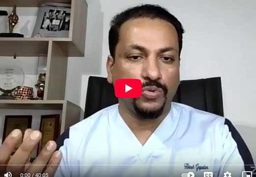
Dr. Garcia’s lecture on meniscus and cartilage transplants
-
Dr Garcia’s Live Lecture on Updated Cartilage Transplants in 2025


Osteochondral Allograft Transplant for Knee Cartilage Repair
Introduction to Osteochondral Allograft Transplantation
Cartilage injuries in the knee can significantly impair function, causing pain, swelling, and mechanical symptoms like catching or locking. Osteochondral allograft transplantation is an advanced cartilage repair option that involves transplanting a section of cartilage and its underlying bone from a donor to repair damaged areas in the knee joint. Unlike other cartilage repair methods, which may require multiple procedures or rely on the patient’s own tissues, osteochondral allografts offer a single-stage solution to restore both cartilage and bone integrity.
This section provides a comprehensive overview of osteochondral allograft transplantation, including its background, indications, required imaging, surgical technique, post-operative care, outcomes, and physical therapy considerations. Designed for patients who wish to understand the procedure and its potential benefits, this guide highlights why osteochondral allografts are a valuable choice for managing extensive cartilage defects in the knee.
Lonzo Ball, cartilage transplant surgery
Cartilage and Meniscus Transplant and Surgical Options
Background on Cartilage Damage and the Need for Allografts
Cartilage, particularly the hyaline cartilage found in knee joints, provides a low-friction, wear-resistant surface that enables smooth joint movement. Once damaged, however, cartilage has a very limited capacity to heal on its own, as it lacks blood vessels, nerves, and lymphatic structures essential for natural repair. When cartilage defects go untreated, they can progress to degenerative changes in the joint, leading to osteoarthritis.
While minor cartilage injuries may be managed with non-surgical methods or smaller procedures like microfracture, larger, full-thickness defects—particularly those with associated subchondral bone loss—often require more substantial intervention. Osteochondral allograft transplantation provides a solution by transferring a section of viable cartilage and subchondral bone from a donor to the patient, effectively addressing both cartilage and bone needs in a single procedure.
Lonzo Ball returns to the 2024 NBA season after cartilage surgery.
Indications for Osteochondral Allograft Transplantation
Osteochondral allograft transplantation is specifically indicated for patients with large, symptomatic cartilage defects in the knee, particularly those that cannot be addressed by other cartilage restoration methods. Key indications include:
1. Large, Full-Thickness Cartilage Defects: Typically, defects larger than 2 cm² in area, especially those involving bone loss, are well-suited to allograft transplantation.
2. Failed Previous Cartilage Repair Surgeries: Patients who have undergone prior cartilage repair techniques (e.g., microfracture or autologous chondrocyte implantation) that did not yield satisfactory results may benefit from an allograft.
3. Osteochondritis Dissecans (OCD): Patients with advanced OCD lesions that involve both cartilage and underlying bone often respond well to allograft transplantation.
4. Young to Middle-Aged Patients: Osteochondral allografts are suitable for younger patients who are too active or too young for total knee replacement and wish to preserve their native joint.
5. Joint Preservation for Athletic Patients: Athletes with cartilage injuries who require significant stability and functionality may find that an allograft offers superior results for a more active lifestyle.
Certain conditions are not typically compatible with osteochondral allograft transplantation, such as widespread osteoarthritis, inflammatory arthritis, or uncontrolled systemic disease, as these can compromise the procedure’s success.
Imaging and Diagnostic Evaluation
A thorough diagnostic workup with imaging is essential to assess the cartilage defect and plan the allograft procedure accurately. Imaging techniques commonly include:
- 1. X-rays: Standard weight-bearing X-rays provide information on joint space, alignment, and any bony abnormalities.
- 2. MRI: An MRI scan is the gold standard for visualizing cartilage defects, evaluating subchondral bone integrity, and identifying other joint structures like ligaments and menisci. MRI also helps in pre-surgical planning by providing an accurate assessment of the defect’s size, location, and depth.
- 3. CT Scan: In complex cases or in patients with prior surgeries, a CT scan may help assess subchondral bone structure and joint alignment. CT can also assist with 3D reconstruction, which is useful for precise graft sizing and placement.
Occasionally, diagnostic arthroscopy may be employed for a definitive assessment. Arthroscopy allows direct visualization of the defect and confirmation of MRI findings, providing the surgeon with additional information to tailor the approach.
Cartilage Repair and Transplantation
Dr. Garcia demonstrates his technique for cartilage transplants for the treatment of knee OCDs
Dr. Garcia demonstrates a cartilage transplant surgery
Surgical Technique for Osteochondral Allograft Transplantation
Osteochondral allograft transplantation involves a meticulous, multi-step process. The procedure is performed in a sterile operating room under anesthesia, typically as an open surgery to ensure accurate placement and fit of the graft.
- 1. Preparation and Sizing of the Allograft: A suitable graft, often from a tissue bank, is matched to the defect size and contour based on preoperative imaging. The graft is taken from a donor and carefully prepared to preserve cartilage and bone integrity. Proper sizing is crucial, as an appropriately fitted graft provides the best biomechanical support and stability.
- 2. Preparation of the Defect Site: The surgeon prepares the defect site by excising damaged cartilage and bone to create a clean, well-defined area for graft insertion. Precise preparation is critical to achieving a stable fit.
- 3. Implantation of the Allograft: The allograft is press-fit into the prepared defect, ensuring stable contact between the graft and the host bone. The graft may be further secured with bioabsorbable screws or pins if needed. The surgeon meticulously aligns the graft to match the surrounding cartilage contour, ensuring a smooth, congruent joint surface.
- 4. Closure: After confirming the stability and alignment of the graft, the incision is closed in layers. Sterile dressings are applied, and a postoperative brace may be fitted to maintain knee stability during initial recovery.
- Osteochondral allograft transplantation is typically completed within 1-2 hours, depending on the complexity of the defect and the graft’s preparation requirements.
Post-Operative Care and Rehabilitation
Post-operative care is critical for graft healing and the success of osteochondral allograft transplantation. The recovery process is carefully structured and often includes the following components:
- 1. Pain Management: Pain control is a priority immediately following surgery. Patients are given oral pain medications, and in some cases, regional anesthesia or nerve blocks are used to reduce pain and enhance comfort.
- 2. Weight-Bearing Restrictions: Weight-bearing is limited to protect the graft. Patients are usually non-weight-bearing on the affected limb for 6-8 weeks, with gradual transition to partial and then full weight-bearing over time.
- 3. Range of Motion (ROM) Exercises: Passive ROM exercises may be initiated within the first few days post-surgery to prevent joint stiffness and aid in healing. Some surgeons recommend a continuous passive motion (CPM) machine to help with gentle joint movement.
- 4. Monitoring for Complications: Follow-up visits are scheduled to monitor healing, check for signs of infection, and assess graft integration. Patients are encouraged to report any symptoms like excessive pain, swelling, or warmth, as these could indicate complications.
3rd time successful patella cartilage transplant with TTO and MQTFL reconstruction
Expected Results and Long-Term Outcomes
Osteochondral allograft transplantation has shown promising outcomes, particularly in younger patients with isolated defects. The success rate can vary depending on factors such as defect size, patient adherence to rehabilitation, and presence of co-existing knee conditions.
Studies suggest that osteochondral allografts offer durable outcomes, with many patients experiencing significant pain relief, increased joint stability, and improved function within 6-12 months post-operatively. Additionally, osteochondral allograft transplants can delay the need for knee replacement by preserving native knee structures.
However, it is essential to note that while many patients experience long-term success, graft survival and function can vary. The presence of risk factors like poor knee alignment, ligament instability, or meniscal deficiency may compromise graft survival.
The Role of Physical Therapy in Recovery
Physical therapy is crucial in achieving optimal outcomes after osteochondral allograft transplantation. A structured rehabilitation program tailored to the patient’s specific needs ensures gradual improvement in strength, flexibility, and joint control.
- 1. Early Range of Motion and Muscle Activation: In the initial stages, physical therapy focuses on gentle ROM exercises and muscle activation without placing excessive load on the graft.
- 2. Gradual Strengthening: Strengthening exercises begin once the graft has had time to integrate, typically around 6-8 weeks post-surgery. Emphasis is placed on strengthening the quadriceps, hamstrings, and gluteal muscles to support the knee joint and reduce stress on the graft.
- 3. Proprioception and Neuromuscular Training: Balance and proprioceptive exercises are essential to improve joint stability and coordination. These are typically introduced during the mid to late stages of rehabilitation.
- 4. Progressive Weight-Bearing and Functional Training: Over time, patients progress to weight-bearing exercises, focusing on proper biomechanics to prevent stress on the graft. For active patients or athletes, sports-specific training is introduced cautiously, usually 9-12 months after surgery.
Conclusion
Osteochondral allograft transplantation offers a valuable treatment option for patients with large cartilage defects who wish to preserve their native knee joint. When performed on well-selected patients, this procedure can alleviate pain, improve joint function, and delay the need for total knee replacement. With careful adherence to post-operative care, rehabilitation, and regular follow-up, osteochondral allograft transplantation can lead to excellent outcomes, enabling patients to return to an active, fulfilling lifestyle.
Dr. Garcia’s technique for patella cartilage transplants with a TTO.
Dr. Garcia’s technique for a larger cartilage transplant called a BioUni. This is a great option to save patients knee’s and reduce arthritis.
Revision cartilage transplant testimonial
HTO and Cartilage Transplant Testimonial
Video testimonial after a revision ACL, meniscus and cartilage transplant in a young athlete


